
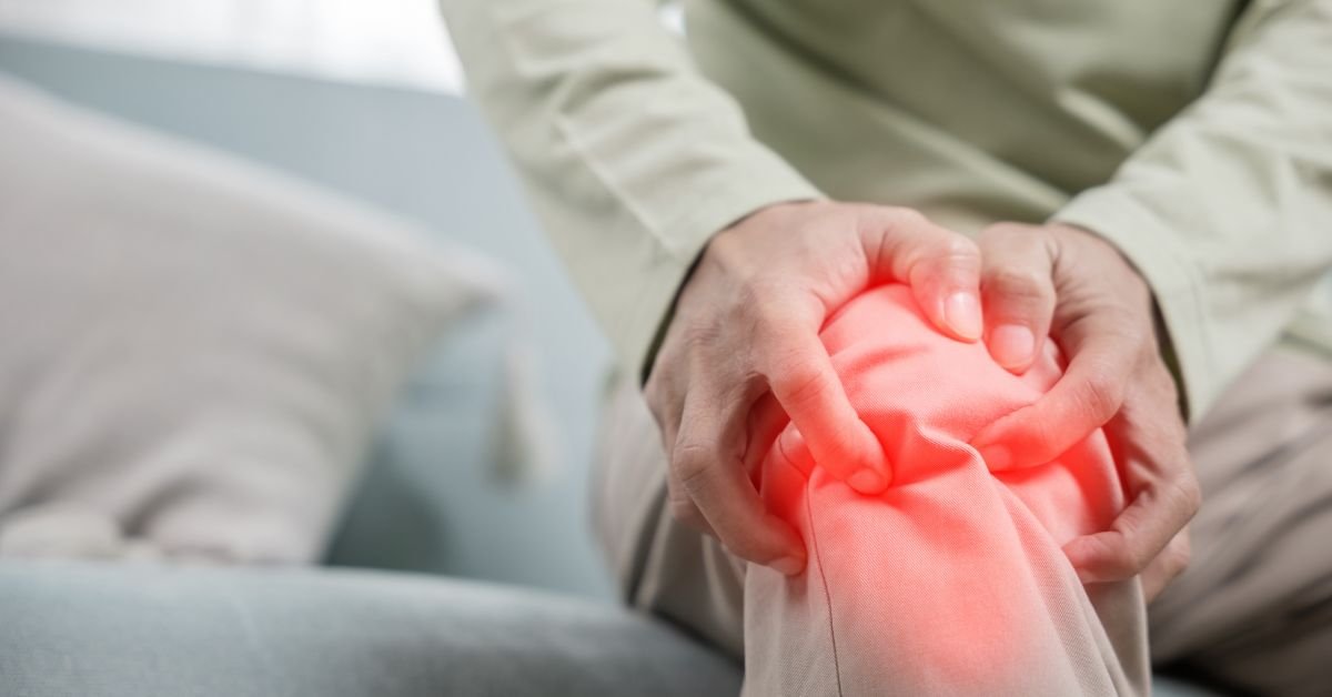
There are a lot of misconceptions about osteoarthritis (OA). Below, we dispel 5 common myths and explain why they aren’t true:
1) OA is caused by wear and tear of the joints and is an inevitable consequence of ageing
Osteoarthritis is known as the classic degenerative age-related joint disease. It has been traditionally understood, by both the general public and health care professionals, as a condition associated with degradation and loss of the articular cartilage, caused by excessive wear and tear of the joints. Its symptoms of joint pain and stiffness have long been accepted as an inevitable part of ageing.
However a considerable amount of new research and understanding challenges this long held myth. It is actually much more complicated. Our joints and bodies are not like car tyres wearing down over time. Our tissues are living cells constantly responding to their environment, including mechanical and chemical stimulation, to maintain homeostasis.
Our joints respond to mechanical and chemical stress by releasing helpful chemicals and proteins to maintain healthy cartilage. What we now understand is that in OA, this homeostasis is lost, and there is a switch to increased production of harmful chemicals and proteins which damage the cartilage. What causes this switch is not yet fully defined or understood, likely because it has different drivers in different individuals.
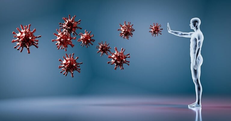
So rather than loss of articular cartilage being driven by ‘wearing out’, OA is better defined as an inflammatory-mediated condition, involving local inflammation, cellular and matrix changes of the cartilage, bone and synovium of the joints. This local joint inflammation, pain, stiffness and swelling is strongly influenced by systemic levels of low grade inflammation, and a wide variety of biopsychosocial factors which have direct and spotify promotion indirect influences on inflammation and tissue sensitivity (1, 2). E.g our lifestyle, diet, activity levels, social environment and beliefs.
Ageing does increase susceptibility: resulting from the effects of ageing on the cells and extracellular matrix of joints, and the wider body system. However increased susceptibility does not mean inevitability. Not all older people develop OA! We must move away from the belief that ageing and excessive use of joints throughout life causes OA. The development of OA and the severity of joint disease and pain is much more closely linked and better explained by other risk factors: genetics, obesity, previous joint injury and certain anatomical factors affecting joint mechanics (3).
To add further weight to this – regular exercise and loading of the joints, bones and muscles is actually protective. Our tissues are living structures which respond and adapt to mechanical load. Numerous studies have shown healthier cartilage, reduced knee pain and lower prevalence of OA in adults who have been physically active throughout their lifetime; including different types of exercise from resistance training to running (4, 5, 6).
Our favourite study demonstrating the beneficial effects of regular exercise is a large systematic review by Alentorn-Geli et al (2017) (7). They analysed data from 25 studies, and 125,000 participants, comparing the prevalence of rates of knee and hip osteoarthritis in runners and non-runners. In non-runners the prevalence was 10.2%. In recreational runners it was a substantially lower 3.5%!
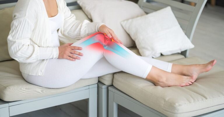
2) Pain correlates with changes seen on x-ray
Radiographic changes observed on x-rays correlate very poorly with osteoarthritis symptoms (8). This might be surprising, but study after study have confirmed this poor correlation. Radiographic features of osteoarthritis can be seen in people with no joint symptoms at all. In addition the severity of symptoms reported by individuals often do not correlate well with the severity of changes seen on an x-ray (9). They are therefore no longer required in best practice guidelines for the diagnosis of OA- the diagnosis can be made clinically.
The poor correlation with changes on x-ray and symptoms can be explained in a number of ways. Radiographic x-rays can detect certain structural changes, traditionally thought of as the hallmark features of OA: such as joint space narrowing and osteophyte formation. However x-rays only provide a snapshot of the joint at one moment in time. They cannot detect sensitised structures. They cannot detect the extremely complicated cell mediated inflammatory processes occurring.
X-rays cannot view the wide range of subchondral and soft tissues structures known to play an important role in the progression and severity of OA disease (cartilage, synovitis, effusion, menisci and ligaments) (10). Furthermore, they cannot tell you anything about the vast range of biopsychosocial influences on that person, their inflammatory response, and their sensitivity at that time. Pain is extremely complicated. You cannot see pain on x-ray
3) Exercise will cause further damage to an osteoarthritic joint
Study after study counter this belief. Numerous studies have followed people with diagnosed and symptomatic OA over a period of time, comparing people who load their joints regularly through exercise, to less active individuals. They have shown that regular loading through exercise is not associated with disease progression, and also perhaps more importantly, is associated with improved self reported symptoms (11, 12).
Exercise and weight loss are the highest recommended treatments for osteoarthritis in best practice guidelines (13, 14). Numerous different types of exercise have been explored in studies, with positive results found for almost any form of exercise. Systematic reviews summarising this evidence have concluded that there is strong evidence supporting the use of aerobic and strength based exercises (land and water) in people with mild and moderate knee and hip osteoarthritis, for improving pain and physical functioning (15, 16).
In addition to improving pain and functioning, strength training has also been shown to alter the progression of the disease at tissue and cellular levels: improved strength, muscle firing patterns and joint biomechanics (17), improved cartilage content (18), and reduced joint space narrowing (19).
The systemic anti-inflammatory and fat reduction benefits of exercise are also huge. Obesity and increased adiposity not only leads to increased load through the joints, the systemic low-grade inflammatory effects are detrimental throughout the body.
Inflammatory products wreak havoc at a cellular level, including on cartilage, muscle tissue, and vast effects on our neuro-immune system leading to increased pain sensitisation (this means our nerves are more sensitive to normal non-threatening stimuli, and our brain is even more sensitive to this information).
4) A joint replacement is inevitable
Disease progression is not inevitable, and joint replacements certainly are not inevitable! In fact individuals show very different trajectories of disease and symptom progression. Long term follow up studies of large populations with knee or hip OA suggest roughly one third of individuals show no radiographic disease progression after 8-15 years (20).
A number of studies have identified prognostic factors for disease progression which include age, BMI, co-morbidity and local joint changes including baseline OA severity, joint effusion and synovitis (21). Interestingly, education level, vitality and pain-coping ability have also been identified as prognostic factors (21).
The average lifetime risk for knee and hip replacements are roughly a third, and a sixth, respectively (20). Individual characteristics do affect this risk, including most notably BMI (20). So in fact the vast majority of people do not go on to require a joint replacement! OA can be very well managed with conservative treatment plans including exercise, weight loss, lifestyle changes and analgesia when required.
5) An increase in pain means the disease is progressing and the joint is getting more damaged
New understanding of pain science tells us that pain correlates very poorly with structural or tissue damage in the body. Pain is extremely complex and multifaceted, and is much better understood as your body’s threat response. Our brains are constantly accumulating a gigantic amount of information from our body to let it know what is going on in the world and how we need to respond.
Pain is an output of the brain, based on large quantities of information including sensations from the body, thoughts, feelings, beliefs, and past experiences. Our brains ‘weigh up’ this information to determine if we are under threat, giving the experience of pain.
Think of pain as your body’s alarm system. Pain is vital for our survival in many situations, i.e. it makes us move our hand away from a flame or stops us from walking on a fractured leg. But sometimes this alarm system is far too sensitive and persists for much longer than needed. Numerous factors can increase the sensitivity of this alarm system including: being anxious, stressed, worried, sleeping poorly, lacking physical activity, lacking social activity, memories about previous pain experiences, previous trauma or psychological distress, as well as genetic and biological factors.
This doesn’t mean pain is ‘in your head’. There are actual chemical and electrical changes occurring in our body’s pain system in response to these including at the level of the nerves, spinal cord and brain itself. These changes make our whole pain/threat/alarm system much more sensitive! Pain is now better explained using a biopsychosocial approach – meaning pain is a complicated and messy expression of biological, psychological and social factors.
This new understanding of pain science goes some way to explaining why individual pain experiences differ so greatly, and why pain is poorly correlated with disease severity or progression in OA. In some cases worsening pain overtime can indicate a progression of disease severity at the local joint level, however in many cases an acute pain flare up or exacerbation, or even gradual worsening of pain overtime, often does not mean there is increasing disease progression or joint damage.
It is much more likely correlated to the numerous other biopsychosocial factors influencing an individual’s pain experience, and can therefore be treated and managed with a holistic approach looking at all of these factors.
The importance of dispelling these myths
Hopefully this post has helped to dispel some commonly held myths about osteoarthritis, which are not only wrong to believe but can also be extremely harmful. For too long people have been considered osteoarthritis as a life sentence of increasing pain and dysfunction, causing them to give up hope and reduce their activity levels to ‘protect’ their joints, leading to a downward spiral of increasing pain, worry, isolation and inactivity.
We are determined to get these new messages out to counteract these beliefs and myths! Please share with article with anybody you feel would find it helpful!
References
- https://www.sciencedirect.com/science/article/pii/S1063458412010254
- https://www.ncbi.nlm.nih.gov/pmc/articles/PMC3638313/
- https://www.ncbi.nlm.nih.gov/pmc/articles/PMC2818253/
- https://research.monash.edu/en/publications/effect-of-physical-activity-on-articular-knee-joint-structures-in
- https://www.aspetar.com/journal/viewarticle.aspx?id=97#.YGr2POhKg2w
- https://www.ncbi.nlm.nih.gov/pmc/articles/PMC2667877/
- https://pubmed.ncbi.nlm.nih.gov/28504066/
- https://pubmed.ncbi.nlm.nih.gov/18764949/
- https://pubmed.ncbi.nlm.nih.gov/15766999/
- https://academic.oup.com/rheumatology/article/57/suppl_4/iv51/4812624
- https://www.ncbi.nlm.nih.gov/pmc/articles/PMC6095814/?fbclid=IwAR0U5wUt95K3spBGXXAZXBieYytC_nAyBmdWHfJBq0Ki2-PGukUx8ffWv6I
- https://www.oarsijournal.com/article/S1063-4584(13)00975-8/fulltext
- https://www.nice.org.uk/guidance/cg177/chapter/1-Recommendations#education-and-self-management-2
- https://pubmed.ncbi.nlm.nih.gov/23595142/
- https://www.ncbi.nlm.nih.gov/pmc/articles/PMC4077018/
- https://www.cochrane.org/CD004376/MUSKEL_exercise-for-osteoarthritis-of-the-knee
- https://www.ncbi.nlm.nih.gov/pmc/articles/PMC3635671/
- https://pubmed.ncbi.nlm.nih.gov/16258919/
- https://pubmed.ncbi.nlm.nih.gov/17013851/
- https://www.sciencedirect.com/science/article/pii/S106345841931088X#bib11
- https://arthritis-research.biomedcentral.com/articles/10.1186/s13075-015-0670-x
